2D ECHO Cardiography
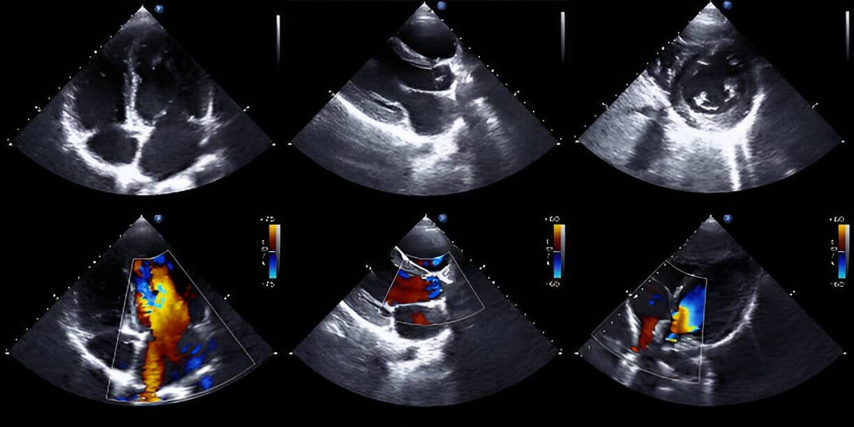
Types of 2D ECHO Cardiography
There are several types of 2D ECHO procedures, depending on your condition and what the doctor needs to evaluate:
Transthoracic Echocardiography (TTE) – The most common type; the probe is placed on the chest to obtain images.
Transesophageal Echocardiography (TEE) – A specialized probe is inserted through the esophagus for clearer images, especially useful when TTE images are inadequate.
Stress Echocardiography – Combines echocardiography with exercise or medication-induced stress to assess heart function under strain.
Dobutamine Stress Echo – Uses a medication (dobutamine) to simulate exercise, typically for patients unable to exercise physically.
Symptoms That May Lead to a 2D ECHO Cardiography
Your doctor may recommend a 2D ECHO if you experience:
Shortness of breath
Chest pain or discomfort
Palpitations or irregular heartbeats
Dizziness or fainting episodes
Unexplained fatigue
High blood pressure or heart murmur detected during a physical exam
What Are Some Common Uses of 2D ECHO Cardiography?
2D ECHO is widely used to:
Diagnose heart valve problems (stenosis or regurgitation)
Assess heart function and pumping ability (ejection fraction)
Detect congenital heart defects
Identify fluid around the heart (pericardial effusion)
Evaluate damage after a heart attack
Monitor known heart conditions and guide treatment decisions
How Do I Prepare for My 2D ECHO Cardiography?
What Will Happen During My 2D ECHO Cardiography?
During the test:
You will lie comfortably on an exam table.
A technician will apply a gel to your chest area to help transmit sound waves.
A handheld device called a transducer will be moved across your chest to capture images.
The procedure is painless, takes about 20–45 minutes, and you can resume normal activities afterward.
For TEE, mild sedation may be given, and the probe will be inserted through the esophagus for clearer imaging.
What Are the Reasons for a 2D ECHO Cardiography?
Doctors recommend a 2D ECHO to:
Investigate symptoms like breathlessness, chest pain, or palpitations
Check heart health in people with hypertension or a history of heart disease
Monitor the progression of heart disease or effectiveness of treatments
Evaluate the heart before and after surgical or medical treatments
Why is 2D ECHO Cardiography Used?
2D ECHO Cardiography is used because it:
Provides detailed, real-time heart images without radiation exposure
Detects early signs of heart problems
Guides treatment planning and follow-up care
Is safe, widely available, and cost-effective compared to more invasive procedures