Elastography
Types of Elastography
There are two main types of elastography:
Ultrasound Elastography (USE)
This method uses sound waves to assess tissue stiffness. It includes:Transient elastography (commonly used for liver)
Shear wave elastography
Strain elastography
Magnetic Resonance Elastography (MRE)
This technique uses MRI to generate detailed images of tissue stiffness, especially useful for deep organs like the liver.
What are Some Common Uses of the Procedure?
Elastography is commonly used to:
Assess liver fibrosis and monitor chronic liver diseases (hepatitis, fatty liver, cirrhosis)
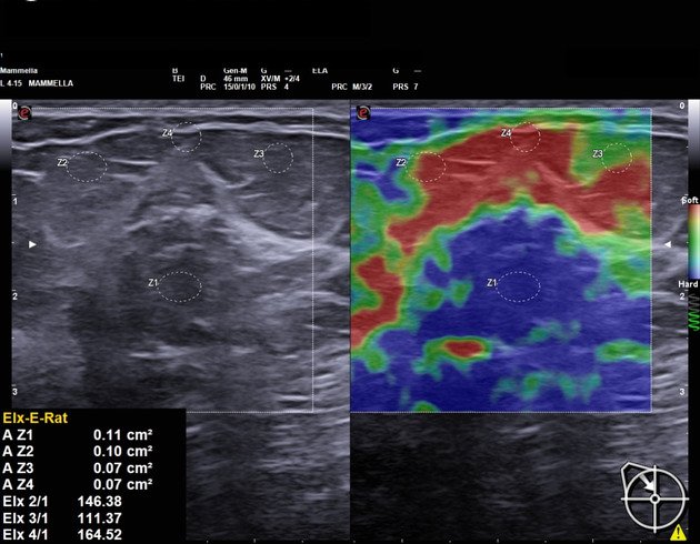
Evaluate breast lumps to differentiate between benign and malignant tumors
Examine thyroid nodules for signs of cancer
Assess prostate stiffness to help detect prostate cancer
Evaluate muscle and tendon injuries
Monitor treatment response in various diseases
How Do I Prepare for My Elastography?
Preparation for elastography is usually simple:
For liver elastography:
Avoid eating or drinking for 2–3 hours before the procedure to improve image quality.
For breast, thyroid, or prostate elastography:
No specific preparation is typically needed.
Wear comfortable, loose-fitting clothes and follow any instructions provided by your doctor.
What Will Happen During My Elastography?
During the scan:
You’ll lie on a cushioned table that slides into the MRI machine
The scan is painless but requires you to lie still for 45–90 minutes
You may hear loud knocking sounds—earplugs or headphones are provided
In contrast scans, a contrast dye may be injected through an IV
Our technician will monitor you throughout and communicate as needed
After the scan, you can resume your normal activities. The images are reviewed by a radiologist, and a detailed report is shared with your referring doctor.
What Are the Reasons for an Elastography?
Doctors may recommend elastography to:
Check for liver scarring in hepatitis B, hepatitis C, or fatty liver disease
Evaluate suspicious lumps in the breast or thyroid
Help differentiate benign from malignant tumors
Avoid or reduce the need for invasive biopsies
Monitor disease progression or treatment response over time