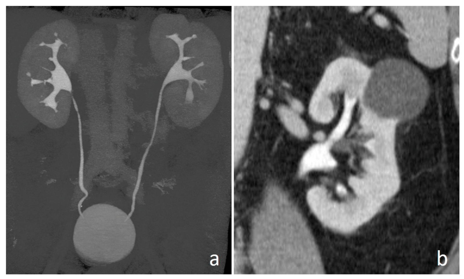
CT Urography / Dual / Triple phase Organ Imaging
CT Urography is a specialized CT scan that provides detailed images of the urinary tract—including the kidneys, ureters, and bladder—using contrast dye to highlight the structures.
Dual Phase and Triple Phase CT Organ Imaging involve taking CT scans at multiple time points after injecting contrast material, capturing how organs like the liver, pancreas, or kidneys absorb and process the contrast. This helps detect tumors, cysts, infections, and vascular abnormalities with remarkable detail.
Types of CT Urography / Dual / Triple Phase Imaging
CT Urography: Focused on the urinary system to detect stones, tumors, structural abnormalities, or blood in urine.
Dual Phase CT: Captures images in two phases (typically arterial and venous) to assess blood flow and tissue enhancement.
Triple Phase CT: Adds a third phase (often delayed) to provide even more detailed evaluation, especially useful in liver and pancreatic imaging.
Common Uses of the Procedure
Evaluate kidney stones, tumors, or infections
Detect urinary tract obstruction or bleeding
Assess liver tumors (hepatocellular carcinoma), hemangiomas, or metastases
Evaluate pancreatic masses, cysts, or inflammation
Examine adrenal gland abnormalities
Map blood vessels before surgery or intervention
How Do I Prepare for My CT Urography / Dual / Triple Phase Scan?
You may be asked to fast for 4–6 hours before the scan.
Inform your doctor if you have kidney problems, diabetes, or allergies to contrast dye.
You may be asked to drink water before the scan to fill your bladder.
Remove jewelry, metal objects, or any accessories near the scan area.
What Will Happen During My CT Urography / Dual / Triple Phase Scan?
You will lie on a comfortable CT table that slides into the scanner.
A contrast dye may be injected into your vein to highlight blood vessels and organs.
The CT scanner will take multiple images, often during specific phases after contrast injection.
The scan is painless, but you may feel a brief warm sensation when the contrast is injected.
The entire procedure usually takes 20–45 minutes, depending on the type of scan.
What Are the Reasons for CT Urography / Dual / Triple Phase Imaging?
Investigating unexplained hematuria (blood in urine)
Detecting tumors or cancers of the kidneys, liver, or pancreas
Evaluating trauma or injury to abdominal organs
Planning for surgery or follow-up after cancer treatment
Assessing vascular abnormalities or aneurysms
Why Are Skeletal and CT Urography / Dual / Triple Phase Imaging Used Together?
In some cases, skeletal imaging (bone scans or CT) may be combined with CT organ imaging to detect bone involvement in cancers, evaluate trauma, or assess conditions that affect both bones and soft tissues. This combined approach gives a comprehensive view of disease spread and helps guide precise treatment.