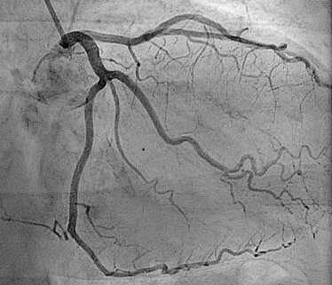
Angiography
Types of Angiography
There are several types of angiography based on the area being examined:
Coronary Angiography – to visualize arteries of the heart
Cerebral Angiography – to examine blood vessels in the brain
Pulmonary Angiography – to check arteries in the lungs
Peripheral Angiography – to assess blood flow in the limbs
Renal Angiography – to examine arteries in the kidneys
CT Angiography (CTA) – non-invasive, using CT scan and contrast
MR Angiography (MRA) – uses MRI technology and contrast dye
What Are Some Common Uses of the Procedure?
Angiography is commonly used to:
Diagnose blocked or narrowed arteries in the heart (coronary artery disease)
Detect aneurysms in the brain or other organs
Evaluate blood flow in stroke or mini-stroke (TIA) cases
Identify blood vessel damage from injuries
Assess conditions before or after vascular surgery
Plan treatment for tumors, especially if they affect blood supply
Monitor blood flow after organ transplants or surgeries
How Do I Prepare for My Angiography?
You may be asked not to eat or drink for 6–8 hours before the test
Inform your doctor if you’re taking medications, especially blood thinners or diabetes drugs
Tell your healthcare provider about allergies, especially to contrast dye, iodine, or shellfish
Wear comfortable clothing and remove any jewelry
You may need to arrange for someone to drive you home after the test
What Will Happen During My Angiography?
The procedure usually takes 30 minutes to 2 hours, depending on the area being examined
You will lie on an X-ray table, and a local anesthetic will be applied
A thin, flexible tube called a catheter will be inserted into an artery (usually in your groin or wrist)
Contrast dye will be injected through the catheter, and X-ray images will be taken
You may feel a warm sensation as the dye flows through your vessels
After the test, pressure will be applied to the insertion site to prevent bleeding
What Are the Reasons for an Angiography?
Doctors recommend angiography when they suspect:
Coronary artery disease (heart blockages)
Brain aneurysms or vascular malformations
Deep vein thrombosis (DVT) or pulmonary embolism
Poor circulation in legs (peripheral artery disease)
Kidney-related vascular issues
Trauma or injury affecting blood vessels
Planning for stent placement, bypass surgery, or angioplasty