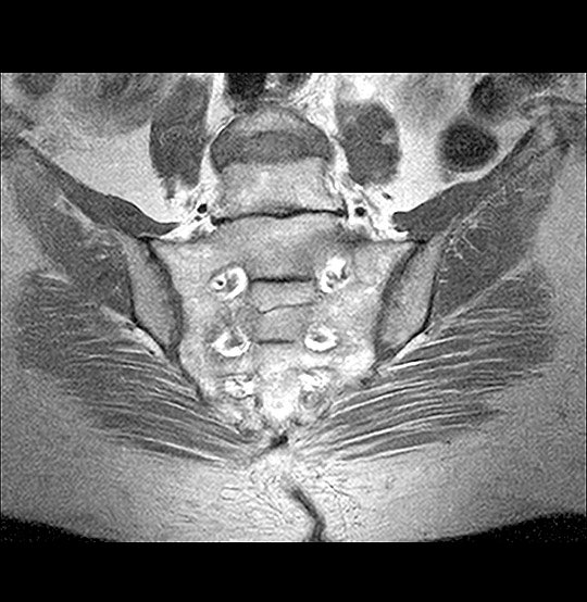
MRI Joints
we offer high-resolution MRI Joint Scans using advanced imaging technology to provide clear and detailed views of bones, cartilage, ligaments, tendons, and muscles—making it easier to detect injury, inflammation, or disease.

What Are Some Common Uses of the Procedure?
MRI Joint scans are commonly used to:
Diagnose ligament or tendon injuries (e.g., ACL tear, rotator cuff tear)
Detect cartilage damage or meniscal tears
Identify joint inflammation, arthritis, or bursitis
Monitor sports injuries or overuse conditions
Evaluate joint infections or fluid buildup
Detect tumors or cysts in or around joints
Guide treatment plans or surgical decisions
Whether you’re an athlete recovering from injury or an individual experiencing chronic joint pain, MRI provides the precision needed for proper evaluation and care.
How Do I Prepare for My MRI Joints?
Preparing for an MRI Joint scan is simple. Most patients do not need any special preparation:
Clothing: Wear loose, comfortable clothes or change into a hospital gown.
Metal Items: Remove jewelry, watches, belts, or any metal objects before the scan.
Medical Devices: Inform your technician if you have any implants, pacemakers, or metal in your body.
Food & Drink: Usually, you can eat and drink as usual unless advised otherwise.
Claustrophobia: Let us know if you are anxious in enclosed spaces—we can offer support or mild sedation if needed.
What Are the Reasons for an MRI Joints?
Doctors recommend MRI Joint scans for various reasons, including:
Unexplained joint pain or swelling
Suspected sports injuries or trauma
Chronic joint conditions like osteoarthritis or rheumatoid arthritis
Evaluation before or after joint surgery
Assessment of mobility issues or functional limitations
Follow-up for abnormal X-rays or physical exam findings
By providing a comprehensive view of the joint’s internal structures, MRI allows for early and accurate diagnosis, which can significantly improve treatment outcomes.
What Will Happen During My MRI Joints?
During the scan:
You’ll lie on a cushioned table that slides into the MRI machine.
The technician will position the affected joint in a comfortable and stable position.
You must remain still while the scanner takes multiple images, which may take 20–45 minutes depending on the joint.
You’ll hear a series of loud tapping or thumping sounds—these are normal.
You can communicate with the technician through an intercom if needed.
The procedure is painless, and most people can resume normal activities immediately afterward.
