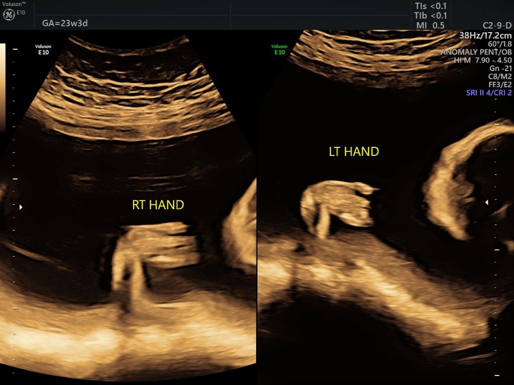
Targetted Obstetric Imaging
Targeted obstetric imaging is a specialized ultrasound scan performed during pregnancy to closely examine the baby’s development, growth, and anatomy. This focused scan allows doctors to evaluate specific fetal structures, the placenta, amniotic fluid, and the uterus to detect any abnormalities or complications early. Unlike a routine pregnancy ultrasound, this targeted scan provides a detailed, in-depth look at specific areas of concern.
we offer Targeted obstetric imaging services to provide expectant parents and doctors with highly detailed, real-time images of the baby and internal organs. This cutting-edge ultrasound technology goes beyond standard 2D scans, delivering clear pictures and live-motion videos, making it a memorable and medically useful experience.

Types of Targeted Obstetric Imaging
Level II Ultrasound (Detailed Fetal Anatomic Survey)
A comprehensive scan to evaluate the baby’s organs, limbs, brain, spine, heart, and other key structures.Fetal Echocardiography
A specialized ultrasound focusing on the baby’s heart to assess heart structure and function.Doppler Ultrasound
Measures blood flow in the umbilical cord, placenta, and fetal vessels, especially useful in high-risk pregnancies.Targeted Growth Scan
Focuses on fetal size, weight, and growth patterns, often done if there’s concern about growth restriction.
Symptoms or Signs That May Lead to Targeted Obstetric Imaging
Your doctor may recommend targeted obstetric imaging if you have:
Abnormal findings on routine ultrasound
Bleeding or pain during pregnancy
History of birth defects or genetic conditions
Abnormal blood test results (such as from first-trimester screening)
Maternal conditions like diabetes or high blood pressure
Multiple pregnancies (twins, triplets)
Common Uses of Targeted Obstetric Imaging
Assess fetal anatomy and rule out birth defects
Check the placenta’s position and function
Monitor amniotic fluid levels
Evaluate fetal growth and well-being
Investigate suspected problems like fetal heart defects, brain abnormalities, or kidney issues
Guide prenatal procedures like amniocentesis
How to Prepare for Targeted Obstetric Imaging
Wear comfortable, loose-fitting clothing.
Drink some water beforehand if advised, as a full bladder may help improve early pregnancy scans.
Bring prior medical reports or previous scan results, if available.
Inform your doctor and the technician about any medications you are taking.
What Will Happen During Targeted Obstetric Imaging
You will lie on a padded examination table.
A gel will be applied to your abdomen to help transmit sound waves.
The sonographer or doctor will use a handheld probe (transducer) to gently move across your belly and capture detailed images.
The procedure usually takes 30 to 60 minutes, depending on the area being examined.
You may be asked to change positions to get clearer views of the baby.
In some cases, a transvaginal probe may be used for better visualization, especially in early pregnancy.
Reasons for a Targeted Obstetric Imaging
Family history or previous pregnancy with birth defects
Abnormal prenatal screening results
Suspected fetal growth restriction or macrosomia (excessive fetal growth)
Maternal medical conditions affecting pregnancy
Multiple gestations (twins or more) requiring close monitoring
